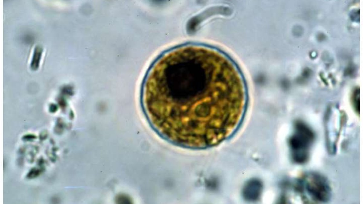
The 2026 Nobel Prize in physiology or medicine has been awarded to three scientists for a discovery that originated with a green alga and a fundamental question about movement. Karl Deisseroth, a psychiatrist and bioengineer at Stanford University, Peter Hegemann of Humboldt University, and Georg Nagel from the University of Würzburg received the honor for revealing how light can regulate brain cell activity. Their work laid the foundation for optogenetics, a technique now used extensively to map brain function.
The research began in a laboratory setting rather than a clinical one. Hegemann and Nagel investigated Chlamydomonas, a single-celled alga commonly found in freshwater ponds, to determine how it detects light. Most living organisms, even those with just one cell, use light to guide their movements—for example, swimming toward food sources or away from harmful conditions. Chlamydomonas accomplishes this through a protein called channelrhodopsin, which is embedded in its cell membrane. When light strikes this protein, its structure changes, creating an opening that allows charged particles such as sodium ions to enter the cell. This influx generates a brief electrical pulse that directs the alga’s movement.
The significance of this discovery became apparent when the researchers realized its potential application in human neurons. Brain cells also rely on electrical signals to communicate. By the early 2000s, Deisseroth had developed a method to introduce the channelrhodopsin gene into brain cells using a harmless viral vector. When exposed to light, these modified cells activate almost instantly, allowing precise control over neural activity.
Mapping fear, memory, and brain circuits
Optogenetics has transformed how scientists study brain circuits linked to memory, movement, and emotional responses. Unlike traditional electrical stimulation, which affects all nearby cells, light can selectively activate only those containing channelrhodopsin. In a 2012 experiment at MIT, researchers conditioned mice to associate a cage with a mild shock. They then used optogenetics to identify and tag the brain cells activated during the fear response. When those same cells were stimulated with light in a different cage, the mice exhibited freezing behavior—indicating a recalled memory of the original shock.
Read Also: Women Speak Out on Unwanted Facial Hair
Since then, the technique has been applied to map brain regions involved in decision-making and addiction. Early-stage clinical trials are also exploring its potential in treating retinal degeneration, where light-sensitive proteins are inserted into surviving retinal cells to partially restore vision. Researchers are also investigating whether optogenetics could eventually be used to treat neurological conditions, including Parkinson’s and epilepsy.
From algae to neuroscience breakthroughs
The initial curiosity-driven research on algae has now evolved into a tool that could transform neuroscience. The Nobel committee emphasized how fundamental scientific inquiry, without immediate practical goals, can lead to new advancements. When Hegemann and Nagel first studied the alga’s light-sensing mechanism, they had no expectation of medical or commercial applications. Yet by exploring how nature functions, they unlocked a method to observe brain activity in real time.
Current efforts focus on expanding optogenetics beyond laboratory settings. While clinical trials for retinal conditions are progressing, challenges remain, particularly in delivering light to deep brain regions without causing tissue damage.
Clinical applications of optogenetics are most advanced in treating retinal degeneration. In ongoing trials, researchers insert genes for light-sensitive proteins, including modified channelrhodopsin, into retinal cells that have lost their natural light detection. Early findings show patients with conditions like retinitis pigmentosa can regain limited vision when exposed to targeted light patterns. The restored cells transmit signals to the brain even when rods and cones are no longer functional.